Hip Muscles Diagram Labeled / Hip Anatomy Pictures Function Problems Treatment : The detailing of these structures changes based on dog breed due to the huge variation of size in dog breeds.
Hip Muscles Diagram Labeled / Hip Anatomy Pictures Function Problems Treatment : The detailing of these structures changes based on dog breed due to the huge variation of size in dog breeds.. The hip muscles encompass many muscles of the hip and thigh whose main function is to act on the thigh at the hip joint and stabilize the pelvis.without them, walking would be impossible. Aaa i gg pp r s t. Large ligaments, tendons, and muscles around the hip joint hold the bones (ball and socket) in place and keep it from dislocating. The piriformis muscle is a key landmark in the gluteal region. For more anatomy content please follow us and visit.
Our latest youtube film is ready to run. Use acronyms to help you. The many muscles of the hip provide movement, strength, and stability to the hip joint and the bones of the hip and thigh. The hamstring muscle group extends across the posterior surface of the thigh from the ischium of the pelvis to the tibia of the lower leg. Ligaments, tendons, and muscles play an important role in the function of the hip.
Muscle Anatomy Of The Hip Anatomy Drawing Diagram from www.med.uio.no They also stabilise the hip joint by 'pulling' the femoral head into the acetabulum of the pelvis. We are pleased to provide you with the picture named labelled diagram of the muscles in the human body.we hope this picture labelled diagram of the muscles in the human body can help you study and research. This diagram depicts hip joint muscles.human anatomy diagrams show internal organs, cells, systems, conditions, symptoms and sickness information and/or tips for healthy living. The detailing of these structures changes based on dog breed due to the huge variation of size in dog breeds. Any injury or disease of the hip will adversely affect the joint's range of motion and ability to bear weight. The largest of them is the most superficial muscle, the gluteus maximus. Understanding the anatomy of the hip is essential for diagnosing its pathology. There are 3 main layers of hip abductor muscles:
Aaa i gg pp r s t.
For more anatomy content please follow us and visit. Muscle, organ and skeletal anatomy). Ligaments are soft tissue structures that connect bones to bones.a joint capsule is a watertight sac that surrounds a joint.in the hip, the joint capsule is formed by a group of three strong ligaments that connect the femoral head to the acetabulum. This diagram depicts hip joint muscles.human anatomy diagrams show internal organs, cells, systems, conditions, symptoms and sickness information and/or tips for healthy living. Just need a glimpse, leave your valuable advice let us know , and subscribe us! Aaa i gg pp r s t. Learn vocabulary, terms and more with flashcards, games and other study tools. The anterior muscle group features muscles. Related posts of muscles of the lower back and hip diagram muscle anatomy amazon. Label the major muscles of the body. They also stabilise the hip joint by 'pulling' the femoral head into the acetabulum of the pelvis. If you're just starting your anatomy journey, work on remembering the names of all 11 hip flexor muscles. As these muscles contract and relax, they move skeletal bones to create movement of the body.
For a more detailed analysis of the muscles used, refer to the biomechanics of rowing. Related posts of muscles of the lower back and hip diagram muscle anatomy amazon. There are 3 main layers of hip abductor muscles: The thighbone or femur and the pelvis join to form the hip joint. The muscles of the pelvis, hip and buttock anatomical chart shows how each muscle in this area of the body works with the others, and the various minor systems within the major ones.
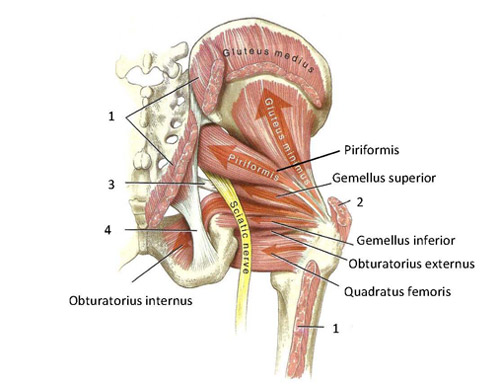
Muscles Of The Gluteal Region 3d Models Video Tutorials Notes Anatomyzone from anatomyzone.com The piriformis muscle is a key landmark in the gluteal region. Included within the chart are gorgeous illustrations of the pelvic diaphragm, sphincter muscles, gluteus maximus muscles, and over a dozen more. The hamstring muscle group extends across the posterior surface of the thigh from the ischium of the pelvis to the tibia of the lower leg. Ligaments are soft tissue structures that connect bones to bones.a joint capsule is a watertight sac that surrounds a joint.in the hip, the joint capsule is formed by a group of three strong ligaments that connect the femoral head to the acetabulum. These muscles can be grouped based upon their location and function. The largest of them is the most superficial muscle, the gluteus maximus. Most modern anatomists define 17 of these muscles. The general action of these muscles is to laterally rotate the lower limb.
Smartdraw includes 1000s of professional healthcare and anatomy chart templates that you can modify and make your own.
Included within the chart are gorgeous illustrations of the pelvic diaphragm, sphincter muscles, gluteus maximus muscles, and over a dozen more. It is also referred to as a ball and socket joint and is surrounded by muscles, ligaments and tendons. Dog anatomy details the various structures of canines (e.g. Our latest youtube film is ready to run. Large ligaments, tendons, and muscles around the hip joint hold the bones (ball and socket) in place and keep it from dislocating. Pick which works for you and then we'll review the muscles! Any injury or disease of the hip will adversely affect the joint's range of motion and ability to bear weight. These muscles can be grouped based upon their location and function. For a more detailed analysis of the muscles used, refer to the biomechanics of rowing. This article will introduce the muscles in each group and touch on their origin, insertion, function, and innervation. This diagram depicts hip joint muscles.human anatomy diagrams show internal organs, cells, systems, conditions, symptoms and sickness information and/or tips for healthy living. The hip muscles encompass many muscles of the hip and thigh whose main function is to act on the thigh at the hip joint and stabilize the pelvis.without them, walking would be impossible. Here are the letters to work with:
The anterior muscle group features muscles. Learn vocabulary, terms and more with flashcards, games and other study tools. The deep gluteal muscles are a set of smaller muscles, located underneath the gluteus minimus. If you're just starting your anatomy journey, work on remembering the names of all 11 hip flexor muscles. Iliopsoas muscle a hip flexor muscle that attaches to the upper thigh bone.
Muscle Diagram Stock Illustrations 4 709 Muscle Diagram Stock Illustrations Vectors Clipart Dreamstime from thumbs.dreamstime.com The alignment and the marrow are the critical elements of the osseous (bony) structures on mr imaging (5). Anatomy of the hip muscles. Here are the letters to work with: The muscles of the pelvis, hip and buttock anatomical chart shows how each muscle in this area of the body works with the others, and the various minor systems within the major ones. Use acronyms to help you. This diagram depicts hip joint muscles.human anatomy diagrams show internal organs, cells, systems, conditions, symptoms and sickness information and/or tips for healthy living. The deep gluteal muscles are a set of smaller muscles, located underneath the gluteus minimus. It helps maintain erect posture, abducts the thigh, and rotates the thigh outward.
If you're just starting your anatomy journey, work on remembering the names of all 11 hip flexor muscles.
Muscle anatomy amazon 12 photos of the muscle anatomy amazon amazon muscle anatomy poster, muscle anatomy amazon, muscle anatomy model amazon, muscle trigger point anatomy amazon, human muscles, amazon muscle anatomy poster, muscle anatomy amazon, muscle anatomy model amazon, muscle trigger point anatomy amazon The thigh bone or femur and the pelvis which is made up of three bones called ilium ischium and pubis. The many muscles of the hip provide movement, strength, and stability to the hip joint and the bones of the hip and thigh. Large ligaments, tendons, and muscles around the hip joint hold the bones (ball and socket) in place and keep it from dislocating. The anterior muscle group features muscles. In human anatomy the muscles of the hip joint are those muscles that cause movement in the hip. Aaa i gg pp r s t. In human anatomy, the muscles of the hip joint are those muscles that cause movement in the hip. They also stabilise the hip joint by 'pulling' the femoral head into the acetabulum of the pelvis. The largest of them is the most superficial muscle, the gluteus maximus. Our latest youtube film is ready to run. Ligaments, tendons, and muscles play an important role in the function of the hip. Attached to the bones of.


0 Komentar